Diagram Of Shoulder - How The Shoulder Works Utah Dr Skedros Orthopaedics. This is the main muscle that lets you rotate and extend your shoulder. The cuff of cartilage increases the surface area of the shoulder joint. Pronate your wrist so the palm of your hand faces down to the floor (as if you were trying to empty a glass of water). The shoulder joint is composed of the glenoid (the shallow shoulder socket) and the head of the upper arm bone known as the humerus (the ball). Bones in shoulder, ligaments of the shoulder joint, parts of the shoulder joint, shoulder anatomy, shoulder joints and muscles, shoulder structure anatomy, shoulder tendon anatomy, shoulder tendons ligaments, human muscles, bones in shoulder, ligaments of the shoulder joint, parts of.
Other important bones in the shoulder include: Four of them are found on the anterior aspect of the shoulder, whereas the rest are located on the shoulder's posterior aspect and in the back. The shoulder also has one articulation, which is the relationship between the scapula (shoulder blade) and the chest wall. The supraspinatus, the infraspinatus, the teres minor and the subscapularis. Its main job is to assist with rotation of the arm away from the body.
Free Shoulders Cliparts Diagram Download Free Shoulders Cliparts Diagram Png Images Free Cliparts On Clipart Library from clipart-library.com Symptoms of rotator cuff tendonitis typically get worse over time. The muscles in the shoulder aid in a wide. The glenohumeral joint is where the ball (humeral head) and the socket (the glenoid) meet. Other important bones in the shoulder include: On the left is a standard (anatomic) shoulder arthroplasty. The shoulder has about eight muscles that attach to the scapula, humerus, and clavicle. The shoulder joint is formed where the humerus (upper arm bone) fits into the scapula (shoulder blade), like a ball and socket. The components of the ball and cup are reversed on the right—a reverse shoulder replacement.
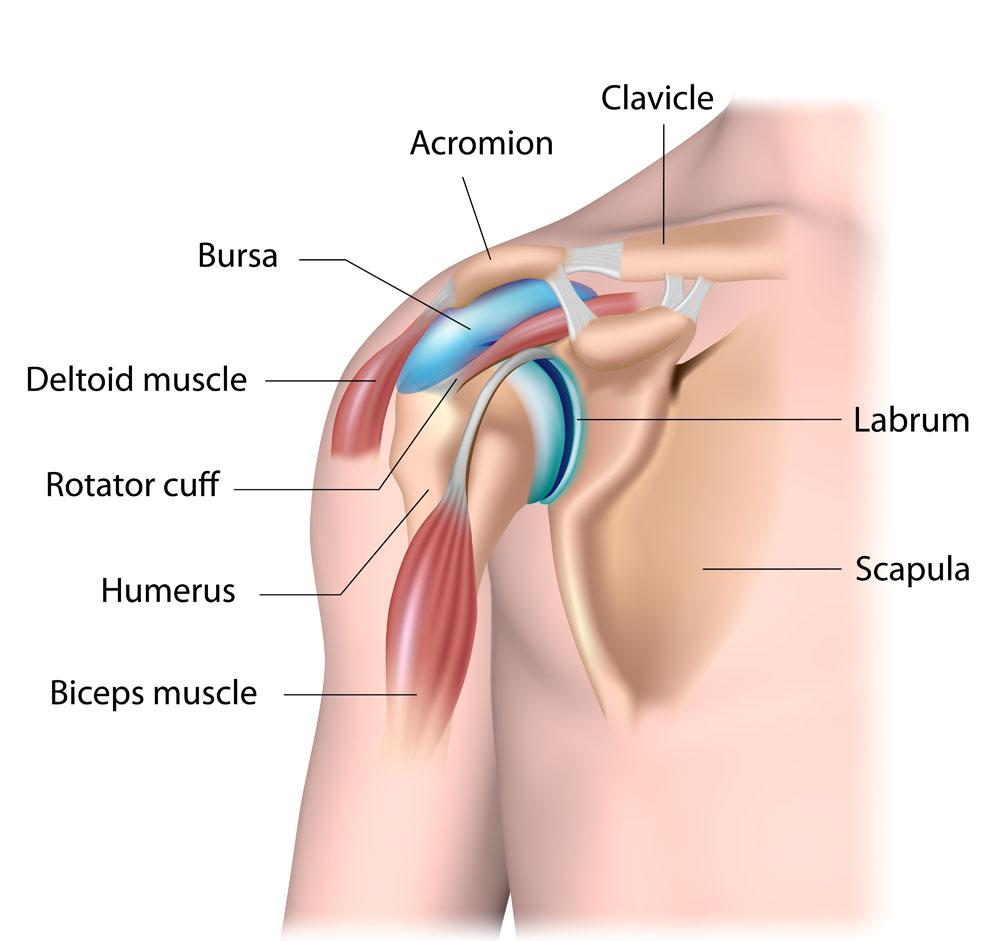
Diagram of the shoulder, including the location of the rotator cuff.
The human shoulder is made up of three bones: The shoulder is made up of two joints, the acromioclavicular joint and the glenohumeral joint. The shoulder has about eight muscles that attach to the scapula, humerus, and clavicle. The shoulder joint can sometimes become narrowed and arthritic, and spurs can form on the undersurface. 17 photos of the diagram of shoulder muscles and tendons. Pain in a man's body pain in a man's body on a gray background. Subscapularis, supraspinatus, infraspinatus and teres minor. The glenohumeral joint connects the shoulder socket, or glenoid, which extends from the shoulder blade, to the arm bone, or humerus. The shoulder joint is the junction between the chest and the upper extremity. A diagram of an anatomic shoulder replacement—the plastic socket replaces the cup of the scapula (shoulder blade). In this condition the rotator cuff is unable to support the glenohumeral joint thereby causing pain in the biceps and the shoulder. The supraspinatus is located on the upper part of the shoulder joint and is involved in abduction (arm raising). These are located in the shoulder blade area, and each related tendon also attaches to the humerus.
A diagram of an anatomic shoulder replacement—the plastic socket replaces the cup of the scapula (shoulder blade). Shoulder tendonitis is inflammation of your rotator cuff or bicep tendons, often caused by overuse of the arms such as in baseball, weight lifting, and racket sports. The shoulder joint is formed where the humerus (upper arm bone) fits into the scapula (shoulder blade), like a ball and socket. On the left is a standard (anatomic) shoulder arthroplasty. There are 10 muscles and 11 shoulder tendons related to shoulder mobility.
Shoulder Anatomy Shoulder Injury Van Nuys Thousand Oaks Los Angeles Ca from lasportsorthomd.com The acromioclavicular (ac) joint connects the upper part of the shoulder blade to the collarbone, or clavicle. In this episode of eorthopodtv, orthopaedic surgeon randale c. A diagram of an anatomic shoulder replacement—the plastic socket replaces the cup of the scapula (shoulder blade). A dislocated shoulder occurs when the humerus (upper arm bone) separates from the shoulder blade at the main shoulder joint. The glenohumeral joint is a joint where the greater tubercle (humeral head at the top of the arm bone) meets the shoulder socket of the scapula, called the glenoid cavity or glenoid fossa. The articulations between the bones of the shoulder make up the shoulder joints.the shoulder joint, also known as the glenohumeral joint, is the major joint of the shoulder, but can more broadly include the. The shoulder socket is a shallow and unstable cavity. The scapula, or 'wingbone', is surrounded by the labrum, allowing the bone of the upper arm (the humerus) to fit into the joint.
Muscles of the shoulder :
The glenohumeral joint connects the shoulder socket, or glenoid, which extends from the shoulder blade, to the arm bone, or humerus. Is the wear and tear of shoulder cartilage until bare bone is exposed. Numerous muscles help stabilize the three joints of. The cuff of cartilage increases the surface area of the shoulder joint. The shoulder joint (glenohumeral joint) is a ball and socket joint between the scapula and the humerus.it is the major joint connecting the upper limb to the trunk. The shoulder joint is protected superiorly by an arch, which is formed by the coracoid process of the scapula, the acromion process of the scapula and the clavicle. All these pictures presented are printable shoulder muscle diagram resources. The glenohumeral joint, the acromioclavicular joint (a/c joint) and the sternoclavicular joint. Treating shoulder pain often takes time but learning how to get rid of shoulder pain through exercise is very simple. The shoulder joint is composed of the glenoid (the shallow shoulder socket) and the head of the upper arm bone known as the humerus (the ball). There are 10 muscles and 11 shoulder tendons related to shoulder mobility. Pain in a man's body pain in a man's body on a gray background. It is one of the most mobile joints in the human body, at the cost of joint stability.
Parts of the right shoulder blade: The muscles of the shoulder support and produce the movements of the shoulder girdle.they attach the appendicular skeleton of the upper limb to the axial skeleton of the trunk. A diagram of an anatomic shoulder replacement—the plastic socket replaces the cup of the scapula (shoulder blade). The acromioclavicular (ac) joint connects the upper part of the shoulder blade to the collarbone, or clavicle. It is one of the most mobile joints in the human body, at the cost of joint stability.
Shoulder Extension Muscles Anatomical Diagram from www.anatomynote.com Sechrest, md narrates an animated tutorial on the basic anatomy of the shoulder. Bones in shoulder, ligaments of the shoulder joint, parts of the shoulder joint, shoulder anatomy, shoulder joints and muscles, shoulder structure anatomy, shoulder tendon anatomy, shoulder tendons ligaments, human muscles, bones in shoulder, ligaments of the shoulder joint, parts of. These are located in the shoulder blade area, and each related tendon also attaches to the humerus. Muscles of the shoulder : The scapula, or 'wingbone', is surrounded by the labrum, allowing the bone of the upper arm (the humerus) to fit into the joint. Symptoms of rotator cuff tendonitis typically get worse over time. Shoulder tendonitis is inflammation of your rotator cuff or bicep tendons, often caused by overuse of the arms such as in baseball, weight lifting, and racket sports. All these pictures presented are printable shoulder muscle diagram resources.
It is one of the most mobile joints in the human body, at the cost of joint stability.
The main shoulder muscles are trapezius, deltoid, pectoralis major and 4 rotator cuff muscles: Here is a list of warmups that will help loosen your shoulder blade muscles and joints prior to exercising. The cartilaginous labrum makes the socket deeper, creating space for the bones to move. This is the smallest rotator cuff muscle. The muscles of the shoulder support and produce the movements of the shoulder girdle.they attach the appendicular skeleton of the upper limb to the axial skeleton of the trunk. The main joint of the shoulder is the glenohumeral joint. The supraspinatus is located on the upper part of the shoulder joint and is involved in abduction (arm raising). Common rotator cuff injuries include rotator cuff tendonitis and rotator cuff strain, which is a partial or complete tear of the rotator cuff. The shoulder joint is composed of the glenoid (the shallow shoulder socket) and the head of the upper arm bone known as the humerus (the ball). The most flexible joint in the entire human body, our shoulder joint is formed by the union of the humerus, the scapula (or shoulder blade), and the clavicle (or collarbone). The muscles in the shoulder aid in a wide. Pain in a man's body pain in a man's body on a gray background. This is the main muscle that lets you rotate and extend your shoulder.
Share :
Post a Comment
for "Diagram Of Shoulder - How The Shoulder Works Utah Dr Skedros Orthopaedics"


Post a Comment for "Diagram Of Shoulder - How The Shoulder Works Utah Dr Skedros Orthopaedics"